Explain the kidney structure in human biology, Biology

Assignment Help:

Explain the Kidney Structure in human biology?

The body's two kidneys are located in the small of the back, one behind the stomach and one behind the liver. Each kidney is composed of a compact outer layer where urine is formed, and a hollow inner sac called the renal pelvis. The renal pelvis is a branched collecting chamber that funnels waste out of the kidney. It also serves as a passageway for the renal artery, which carries blood to the kidney, and the renal vein, which carries away filtered blood.

1875_kidney.png

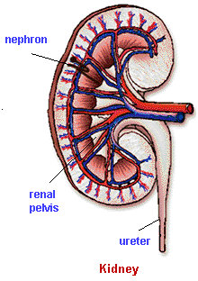
The outer layer of the kidney is divided into the renal medulla and the overlying renal cortex. Structures called nephrons are found in the cortex and extend into the medulla. The kidney contains approximately one million nephrons, which carry out the major functions of the urinary system.

An arteriole supplies each nephron with blood, and a venule drains it. The arteriole runs into a capillary bed called a glomerulus, which is enclosed in a cup-shaped structure called Bowman's capsule. The glomerulus and its surrounding Bowman's capsule compose the renal corpuscle.

The renal tubule is divided into three segments. The proximal convoluted tubule, located in the cortex, leads to a tubule segment known as the loop of Henle as it extends down into the medulla, where it makes a hairpin loop before returning to the cortex. At this point it becomes the distal convoluted tubule, so called because it is away from, or distal, to the glomerulus. Several tubules join a collecting duct, which passes through the medulla adjacent to the loop of Henle.


Related Discussions:- Explain the kidney structure in human biology

Describe new cardio-vascular risk factors, Describe New Cardio-vascular Ris...

Describe New Cardio-vascular Risk Factors ? The major risk factors contributing to the development of atherosclerotic plaques in blood vessels have been known for many years. H

Chromosomes with spindle fiber, Chromosomes with spindle fiber: The kineto...

Chromosomes with spindle fiber: The kinetochore fibers of spindle attach to the kinetochore region (specialized area in centromere) of chromosome, and align them at the equator of

Karyokinesis, At the beginning of the process in an animal cell, the partit...

At the beginning of the process in an animal cell, the partition of the centriole takes place, which has been duplicated during Interphase but were in the same centrosome. Bipol

Pre-embryonic development, Pre-Embryonic Development We had said in t...

Pre-Embryonic Development We had said in the starting of the unit that prenatal development can be divided into two phases, embryonic and foetal. The embryonic phase of devel

What is coronary artery disease, Q. What is Coronary artery disease? Nu...

Q. What is Coronary artery disease? Nutrition is a basic prerequisite for maintaining a desirable body weight and that diet has a major role in the prevention and treatment of

What are the problems that vertebrates required, What are the problems that...

What are the problems that vertebrates required to solve to adapt to the terrestrial environment as they came from the aquatic habitat? How evolution does solve those problems?

Explain the term - social attention and environmental influe, Explain the t...

Explain the term - Social Attention and Environmental Influences The importance of social environment and early experiences in neurodevelopment is increasingly recognised. Due

Chemical and heat burn, Chemical and Heat Burn Chemical born and Heat ...

Chemical and Heat Burn Chemical born and Heat burn injuries have currently started receiving greater attention. The reason for this is perhaps the degree of discomfort and per

Heparinisation, Heparinisation Blood when allowed to flow out from ...

Heparinisation Blood when allowed to flow out from body to the circuit tubings can get clotted. To prevent this, heparinisation is done. Patient is administered 3 mg

What is resting membrane potential of an axon, Action potentials include ch...

Action potentials include changes in the membrane potential of an axon, as shown in the schematic below. During the refractory period, sodium channels are inactivated and

Write Your Message!

Captcha
Free Assignment Quote

Assured A++ Grade

Get guaranteed satisfaction & time on delivery in every assignment order you paid with us! We ensure premium quality solution document along with free turntin report!

All rights reserved! Copyrights ©2019-2020 ExpertsMind IT Educational Pvt Ltd